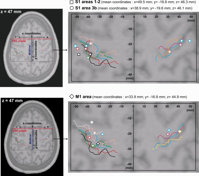
Figure 1.

Localization of intracerebral contacts exploring the pre‐ and postcentral gyri. The position of each contact in a given patient was determined in three dimensions using his/her own 3D‐MRI. Then, positions of all contacts were registered in the Talairach and Tournoux referential and plotted together on a horizontal MRI slice, with z coordinate corresponding to the mean z coordinate of contacts in S1/M1 (z = 47 mm). Colored lines illustrate the position of the central sulcus in each patient. The same color was used to represent the central sulcus and the electrode contact positions in a given subject. Contacts located in S1 areas 1–2 are represented by squares, contacts located in S1 area 3b by circles and contacts located in M1 by diamonds. All the contacts are located according to their x (distance between each contact and the midline plane) and y coordinates (distance between each contact and the Vertical Anterior Commissure (VAC) plane).
