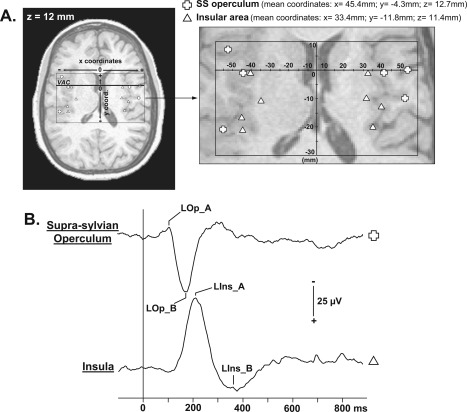
Figure 2.

Localization of contacts exploring supra‐sylvian opercular and insular cortices and LEPs recorded in these areas. A: According to the same procedure described in Figure 1, positions of all contacts recorded in operculum (open cross) and insula (open triangle) are represented on a horizontal MRI slice (z = 12 mm). B: Supra‐sylvian opercular and insular LEPs grand average from all the patients (referential recording mode; positivity: downward). VAC: Vertical Anterior Commissure plane. SS operculum: Supra‐sylvian operculum.
