
Figure 1.
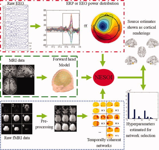
Schematic representation of NESOI. The raw EEG data is processed for artifact‐rejection and the amplitude and/or other features of interest are extracted. The corresponding fMRI data are pre‐processed and separated into spatially independent components. The structural MRI is segmented to provide a forward model for EEG imaging. The intensity of the neural electric sources and the hyperparameters are iteratively estimated by NESOI.