
Figure 1.
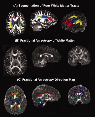
White matter tract segmentation and fractional anisotropy (FA) quality (A) Axial and sagittal magnetic resonance images showing the segmentation of four white matter tracts [red = Anterior Corona Radiata; green = Posterior Corona Radiata; blue = SCR; yellow = Superior Longitudinal Fasciculus] and a three dimensional transparent head image with segmented white matter tracts. B: White matter FA shown in axial, coronal, and sagittal planes. C: White matter FA direction, depicted in color [red = commissural (left‐right) pathways; cyan = association (Anterior‐Posterior) pathways; purple = projection (Inferior‐Superior) pathways], fused with mean diffusivity axial, coronal, and sagittal images. FA is modulated by the principal eigenvector to produce the color map and this color map is fused with the mean diffusivity. This fused map provides a clear depiction of gray matter, white matter, and cerebrospinal fluid.