
Figure 6.
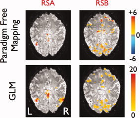
Statistical maps obtained with PFM (P value < 0.05, FDR corrected) (top) and GLM (F‐test, P value < 0.05, FDR‐corrected) (bottom) corresponding to the time points of activations shown at the ATS of subject A at TR 0.4 in Figure 2. [Color figure can be viewed in the online issue, which is available at wileyonlinelibrary.com.]