
Figure 1.
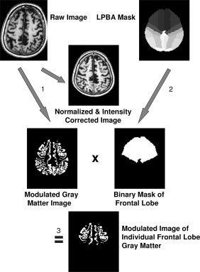
Image processing and volume determination shown exemplarily for the frontal lobe gray matter: (1) Unified segmentation of SPM5 (i.e., normalization, segmentation, and intensity correction) is performed on a T1‐weighted volume data set. (2) A binary frontal lobe mask is derived from the LONI Probabilistic Brain Atlas (LPBA40) by setting all voxels of the maximum likelihood map belonging to the frontal lobe to a value of “one” while all other voxels are set to zero. (3) The modulated gray matter image resulting from unified segmentation is multiplied with the frontal lobe mask. This results in a modulated image of the individual frontal lobe gray matter. Because of modulation of the gray matter image, the effect of normalization (i.e., extension or shrinkage of the investigated structure) is compensated for so that the computed volume represents the volume of the original structure in native space. The analysis is fully automated and requires about 1 h per MRI scan on an AMD Opteron 2.0 GHz PC.