
Figure 2.
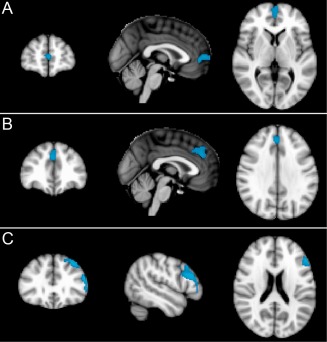
ASL: Areas of significantly (cluster level corrected P < 0.05) reduced regional cerebral blood flow (rCBF) in older relative to younger participants in three clusters in the frontal lobes in the midline and left dorso‐lateral frontal regions. Slides in radiological convention with left hemisphere to the right of the image.