
Figure 2.
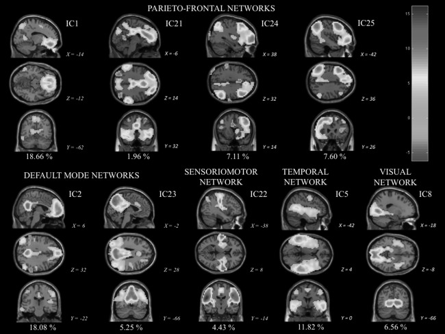
Visually identified group‐level components obtained by temporal concatenation group independent component analysis (TC‐GICA): fourth parietofrontal networks (IC1, IC21, IC24, and IC25), two default mode networks (IC2; IC23 = posterior cingulate/ precuneus subsytem), one sensoriomotor (IC22) network, one mainly temporal network (IC5), and one visual network (IC8). Sagittal, coronal, and axial views (showed as x, y, z according to MNI152 standard space) of these nine functionally relevant group‐level independent components (ICs) are displayed according to neurological convention (right on right). The percentage of variance explained by each IC is displayed below each IC. The z‐statistic map legend for all networks is shown in the up right corner. [Color figure can be viewed in the online issue, which is available at http://wileyonlinelibrary.com.]