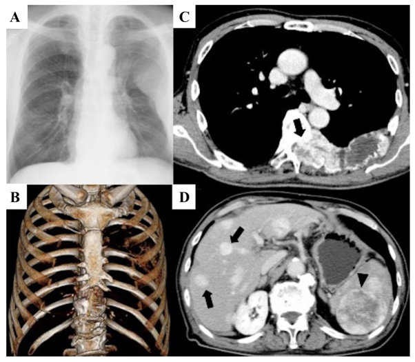
Figure 1.
Large osteolytic lesions were confirmed in the 6, 7 and 8th left ribs and in the 6 and 7th thoracic vertebra. (A) Roentgenogram of the chest. (B) Three-dimensional CT. (C) CECT scan of the thorax showing that the spinal canal was occupied by these lesions, and the lesions were well-contrasted (arrow). (D) CECT scan of the abdominal region showing that massive lesions were found in the liver (arrow) and spleen (arrow head). CECT, Contrast-enhanced CT.

