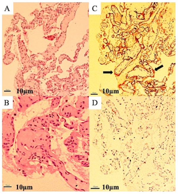
Figure 3.
Microscopic examination. (A) Low-powered microscopic examination showing a replacement of bone tissue with vascular tissue and a netlike proliferation of blood vessels similar to capillary vessels consisting of monolayered vascular endothelium (magnification, ×40). (B) High-powered microscopic examination showing hyaline fibrous interstitium. No cellular atypia is present, and osteoblastic response is absent (magnification, ×100; scale, 10 µm). (C) Immunohistochemical examination showing an overexpression of cluster of differentiation-34 (arrow) on vascular endothelial cells (magnification, ×40). (D) The MIB1 proliferation index ranged from 8 to 10% (magnification, ×40). The MIB-1 index was calculated by counting positive and negative nuclei at any 10 hot spots after immunostaining and calculating the ratio visually.

