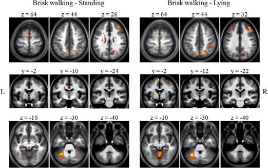
Figure 2.

Comparison of BOLD signal changes in fMRI during mental imagery of brisk walking compared with mental imagery of standing and mental imagery of lying. During imagination of brisk walking compared with imagination of standing and lying prominent activations were found in fronto‐parietal regions, basal ganglia, posterior cingulate cortex, anterior insula, left parahippocampal gyrus, and cerebellum. Results are displayed on the group mean high resolution T1 weighted MRI at a threshold of P < 0.05, FDR‐corrected.
