
Figure 2.
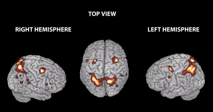
Brain regions activated by incongruence (Incongruent > Congruent trials). Clusters showing higher activity in the incongruent than congruent condition irrespective of distracter and effector are rendered on three‐dimensional (3D) views of the SPM template. This contrast revealed the activation of frontal and parietal regions. The frontal region included the left Precentral Gyrus (L Precentral G), the right Middle Frontal (L/R FEF) cortex bilaterally, the right Supplementary Motor Area (R SMA), the most posterior portion of the Inferior Frontal Gyrus, the Operculum, bilaterally, and the Pars Triangularis (IFG) extending into the Insula and the middle portion of the right Cingulate Cortex. The parietal region included the right superior and inferior Parietal Cortex bilaterally. These regions were used as ROI to assess any differential influence of distracter/instruction signal incongruence on brain responses (SPM thresholds are set to P corr. = 0.05 at cluster level).