
Figure 2.
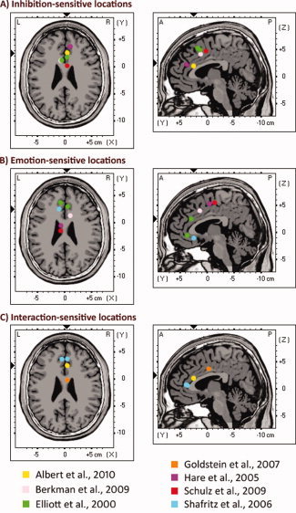
Depiction of location of MNI coordinates used to define the regions of interest (ROIs) within the ACC and the functionally related areas of the medial wall. For presentation purposes, coordinates were collapsed on a representative brain slice at Z = 25 (axial view) and Y = 4 (sagittal view). Exact coordinates are given in Table III. These coordinates represent the center of the ROIs (radius = 9 mm). All these locations have been shown to be activated in previous studies on emotional response inhibition. A: ACC locations previously associated with response inhibition. B: ACC locations previously associated with emotional processing. C: ACC locations previously associated with the interaction of emotional processing and response inhibition. An interactive animation reproducing the location of each MNI coordinate employed to define the ROIs (projected one by one on sagittal, axial, and coronal slices of the Colin brain) can be seen at http://www.uam.es/carretie/grupo/cooACC.htm. [Color figure can be viewed in the online issue, which is available at wileyonlinelibrary.com.]