
Figure 3.
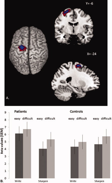
Differential cerebral effects of imagined writing in patients and controls. Panel A shows cerebral activity that increased during motor imagery of writing vs. motor imagery of sharpening, and more in patients than in controls. This contrast is shown at an uncorrected threshold of P < 0.05 and P < 0.01 (for graphical purposes), overlaid onto the structural scan of a representative subject from the MNI series. To restrict these effects to areas specifically involved in motor imagery, we included only voxels where activity increased linearly with repetition (2, 4, or 6 imagined movements) and where activity increased more for biomechanically difficult than easy trials (using inclusive masking at an uncorrected threshold of P < 0.05). The left side of the figure is the left side of the brain. Panel B shows cerebral responses over the left dorsal premotor cortex (PMd; −24 −6 +66]. This region showed a significant Group × Task interaction (P = 0.048, FDR‐corrected). For each group, the histograms show parameter estimates (in S.E.M. units) evoked by writing (the left two bars for each group) or sharpening (the right two bars for each group), separately for trials involving biomechanically easy movements (dark gray bars) or biomechanically difficult movements (light gray bars). Effects are averaged over the different repetitions (2, 4, or 6 imagined movements). [Color figure can be viewed in the online issue, which is available at wileyonlinelibrary.com.]