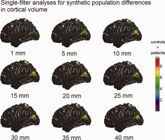
Figure 4.

Synthetic population differences in cortical volume (t maps, RFT thresholded with tα = 0.05) detected with single‐filter analyses. The color bar encodes the t‐statistics at each vertex. Bellow each t map is the corresponding filter size. [Color figure can be viewed in the online issue, which is available at http://wileyonlinelibrary.com.]
