
Figure 1.
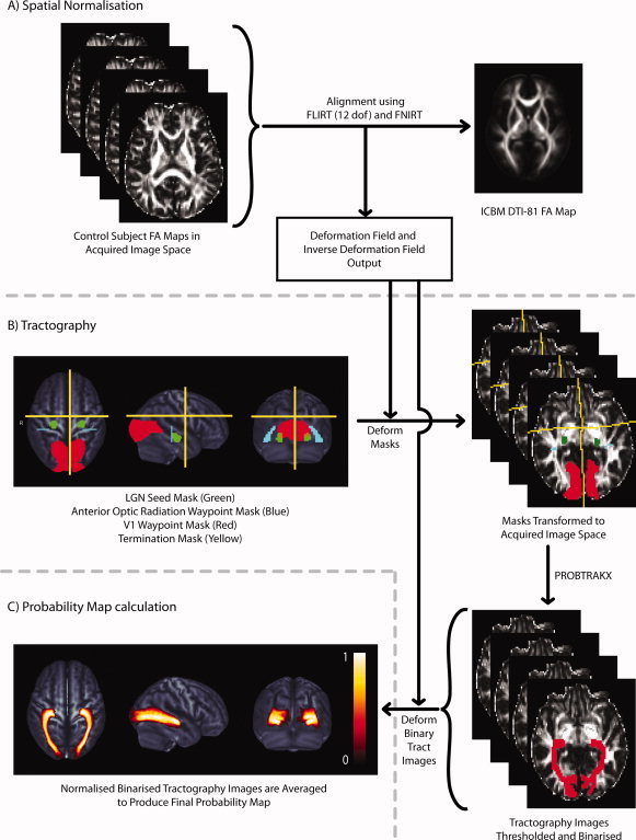
Illustrates the methodology used to create the probability map of the optic radiations. (A) Each subject's FA image was registered to the ICBM template FA image using a two‐step registration procedure involving an initial linear alignment followed by a nonlinear deformation. The deformation field and inverse deformation field were output. (B) Seed masks in the lateral geniculate nuclei (LGN, green), waypoint masks in the anterior optic radiation (blue) and primary visual cortex (V1, red) and termination masks used to remove spurious interhemispheric, anterior and superior projecting streamlines (yellow) were defined in standard space, deformed to each subject's DW image space and used for probabilistic tractography. (C) Individual subject binary tract images were deformed to standard space and averaged to created the probability map which is displayed here as an orthogonal maximum intensity projection. [Color figure can be viewed in the online issue, which is available at wileyonlinelibrary.com.]