
Figure 1.
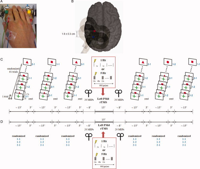
A: Right task hand of one subject resting in pronated position on the test table. Surface electromyography was recorded simultaneously from first dorsal interosseus (FDI) and abductor digiti minimi (ADM) muscles, the primary movers of index finger and little finger abductions, respectively. The in‐house manufactured device for recording button presses by index finger abduction (green button, 1) and little finger abduction (yellow button, 2) triggered automatic marker events in the EEG recording system. B: Sites of focal TMS (figure‐of‐eight coil indicated) over the hand area of the left primary motor cortex (M1, blue spot) and the left dorsal premotor cortex (PMd, red spot) shown on a 3D MRI reconstruction of one individual brain. The site of PMd stimulation was targeted by MR‐navigation (see Methods) and was located, on average, 1.8 ± 0.3 cm anterior of the M1 stimulation site. The M1 site was used for testing corticospinal excitability, the PMd site was used for application of rTMS (see below). C: Experimental protocol for the contingent negative variation (CNV) sessions. Subjects performed 3 × 81 CNV trials before and after left PMd‐rTMS (either 1 Hz or 5 Hz rTMS in separate sessions). Each CNV trial consisted of a warning stimulus (green cross) followed 2 s later by an imperative go‐signal (all signals presented on a screen in front of the subjects) instructing on the motor response to be performed (red square: index finger abduction repeated two times; red circle: index finger abduction followed by little finger abduction; red triangle: little finger abduction followed by index finger abduction). The order of go‐signal conditions was pseudo‐randomized. The interval from the go‐signal to the warning signal of the next trial varied between 3–7 s. Corticospinal excitability of the hand area of the left M1 was tested immediately before and after PMd‐rTMS by recording 20 MEPs in the resting FDI of the right‐hand. D: Experimental protocol for the Bereitschaftspotential (BP) sessions. This protocol were identical to the CNV protocol, except that the 2‐item right hand finger abduction task (same movement conditions as in the CNV trials) was performed self‐initiated without external cuing (∼1 trial every 7 s). The subjects had to select internally one of three motor conditions for the upcoming next trial and were instructed to randomize these conditions over the 3 × 81 trials before and after left PMd‐rTMS.