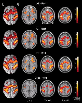
Figure 6.

Across‐subject (n = 23) conjunction activation for IAT–Rest, AT–Rest, PT–Rest and MSC–Rest. Areas of significant activation are overlaid on normalized brain slices based on the anatomic T 1 slices. The main areas of activation for the IAT, AT and PT tasks are seen bilaterally in the SI, SII, LOC, SMA, PFC, IPS and left MI regions, but no activation is seen in right MI regions. In contrast, the main areas of activation for the MSC task are seen bilaterally in the SI, SII, PFC and left MI regions, but not in the LOC, SMA and IPS regions, which are associated with higher‐level tactile angle discrimination. Note the absence of primary visual cortex activation. L, left side; R, right side. [Color figure can be viewed in the online issue, which is available at wileyonlinelibrary.com.]
