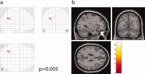
Figure 2.

Increased MD in ET patients compared to control is detected in white matter adjacent to the left parieto‐occipital sulcus (P = 0.005). Visualisation as SPM “glass brain” maximum intensity projection (a), and overlaid onto a canonical single subject brain (b).
