
Figure 3.
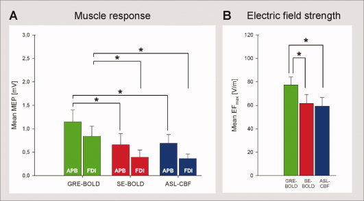
Stimulation of brain tissue at fMRI peak voxel coordinates with TMS. Neuronavigated TMS (120% resting motor threshold) was used to stimulate brain tissue at fMRI peak voxel coordinates to investigate whether localization differences between fMRI sequences are functionally relevant, i.e., impact on muscle responses (n = 12 subjects). A: Muscle responses. Peak‐to‐peak motor‐evoked potentials (MEPs) of the abductor pollicis brevis (APB) and the first dorsal interosseus (FDI) hand muscle are shown. MEPs resulting of stimulation at GRE‐BOLD coordinates were significantly higher than MEPs resulting of stimulation at SE‐BOLD and ASL‐CBF coordinates. B: Electric field strength at target position. Maximum electric field values (EFmax) reaching the fMRI target position during stimulation are shown. GRE‐BOLD coordinates were stimulated with significantly higher EFmax values than SE‐BOLD and ASL‐CBF coordinates which were significantly deeper in the central sulcus and hence further away from the TMS coil. Mean EFmax values during stimulation at SE‐BOLD and ASL‐CBF coordinates were not statistically different (paired‐sample t‐test, P > 0.05). Error bars indicate standard errors. Asterisks indicate significant differences in paired sample t‐tests (paired‐sample t‐test, P < 0.05, each comparison).