
Figure 1.
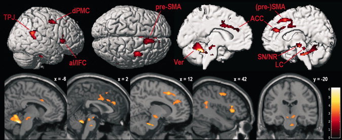
Supramodal alertness‐related brain activity (averaged unimodal alertness conditions vs. averaged control conditions, masked to only include voxels that showed stronger activity during each unimodal alertness condition than during resting baseline and the respective control condition). TPJ, temporo‐parietal junction; dPMC, dorsal premotor cortex; aI/IFC, anterior insula/inferior frontal cortex; pre‐SMA, pre‐supplementary motor area; Ver, cerebellar vermis; ACC, anterior cingulate cortex; SN/NR, substantia nigra/nucleus ruber; LC, locus coeruleus. Parasagittal slices show activity overlaid over the SPM5 single‐subject template brain; coordinates refer to MNI space; color codes t values; voxel‐wise P < 0.001 and FWE‐corrected cluster‐level P < 0.05.