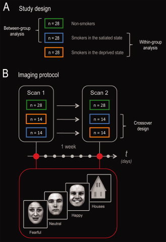
Figure 1.

Study design and imaging protocol. (A) Smokers (and nonsmokers) were scanned on two separate occasions 1 week apart: smoking satiety versus overnight deprivation, in a randomized counterbalanced order. This enabled us to perform both between‐group (nonsmokers vs. satiated smokers) and within‐group (satiated state vs. deprived state) analyses. (B) To specifically assess amygdala function, smokers and nonsmokers were scanned on a face perception task including fearful, happy, and neutral facial expressions (plus houses as nonfacial control stimuli).
