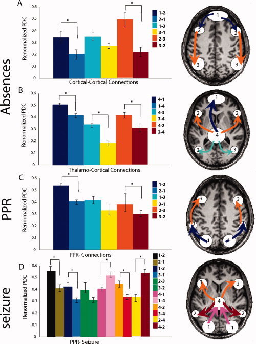
Figure 3.

Renormalized directed partial coherence (RPDC). In the left of the figures RPDC for all directions of connections are shown in bar graphs. Directions which were significantly stronger are indicated by an asterisk. In the right RPDC connections which were significantly stronger are shown schematically for all sources overlaid on the brain (A) cortical–cortical connections of the sources in absences: information flow from the frontomedial cortex via the prefrontal cortex to the parietal cortex (B) thalamo‐cortical connections of the sources in absences: RPDC from the thalamus to all other sources are stronger. (C) cortical connections of the sources in PPR: information flow from the occipital cortex via the intraparietal slucus to the premortor cortex (D) cortical–cortical and thalamo‐cortical connections of the sources in a patient in whom PPR was followed by a generalized tonic‐clonic seizure.
