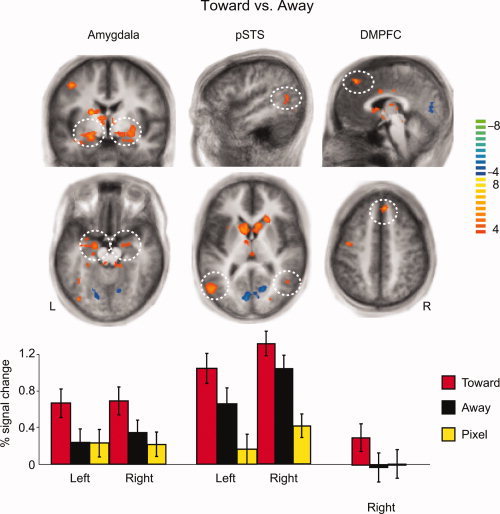
Figure 3.

Different response patterns of the amygdala, pSTS and dmPFC in the Toward, Away, and Pixels conditions. Top and middle panels: The contrast of Toward vs. Away was associated with brain activation in the amygdala, pSTS, and right dmPFC. Bottom panel: The signal changes within the above areas show that the amygdala response was stronger in the Toward condition than either in the Away or Pixel conditions, whereas the pSTS showed enhanced responses to both types of human stimuli compared with the pixel images, and the dmPFC showed activation only in the Toward condition. The volumes obtained from the contrast are 888 mm3 area in left amygdala, 309 mm3 area in right amygdala, 1,678 mm3 area in left pSTS, 348 mm3 area in right pSTS, 629 mm3 area in right dmPFC; the color bar on the right represents the t‐values, and the axial slices are from the Talairach z‐coordinates of −14, 7, and 44 respectively from left to right.
