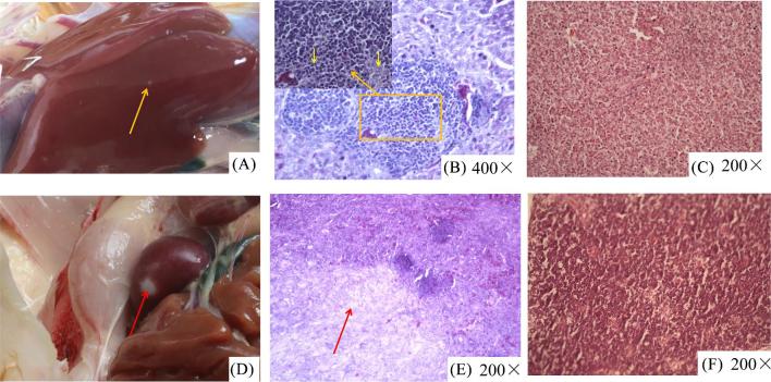
Figure 6.
Anatomical and histological lesions. (A) White tumor nodules, approximately 2 to 3 mm in diameter, on the liver surface. (B) Lymphocytic proliferation foci were found in the livers. (C) Normal liver tissues. (D) Sarcoma in the spleens. (E) A large number of irregular fibrous cells infiltrated into spleen tissues. (F) Normal spleen tissues.

