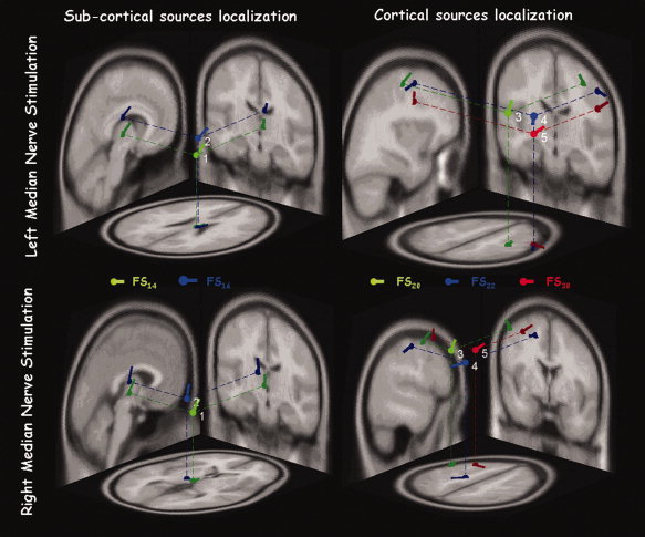
Figure 4.

Single subject FS positions. Position, direction, and orientation of the ECD corresponding to each FS in response to the left and right separate median nerve stimulation, in the MNI brain template––axial, coronal, and sagittal views. [Color figure can be viewed in the online issue, which is available at www.interscience.wiley.com.]
