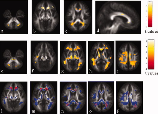
Figure 2.

Top row: statistical parametric mapping (SPM) regions (color‐coded for t values) with decreased white matter (WM) fractional anisotropy (FA) values in patients with primary progressive multiple sclerosis (PPMS) compared with controls (P < 0.001, FDR corrected). Several regions in the frontal and temporal lobes, the CC, the cingulum, the OR, and the middle cerebellar peduncle, bilaterally, appear to be damaged. Middle row: SPM regions (color‐coded for t values) with increased WM mean diffusivity (MD) in PPMS patients compared with control subjects (P < 0.001, FDR corrected). Widespread MD abnormalities in the major short and long intra‐hemispheric associative pathways, as well in the CC and cingulum are visible. Bottom row: SPM regions with anatomical correspondence between decreased WM FA (red) and increased WM MD (blue). An overlap is visible in the CC, the cingulate gyrus, the left short temporal fibers, the right short frontal fibers, the OR, bilaterally, and the middle cerebellar peduncles. Images are in neurological convention. See text for further details.
