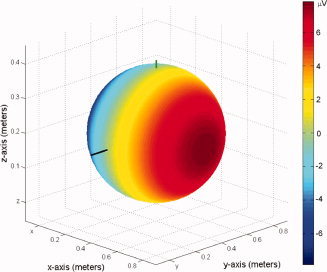
Figure 8.

Analytic simulation of surface potentials generated at 3 T by flowing blood in the posterior‐to‐anterior direction through a vessel modeled after the internal carotid artery, given a 19‐cm sphere, cylindrical flow of 0.5 cm in diameter and 1 cm in length, and flow velocity of 12.6 cm/s in the posterior‐to‐anterior direction. Note that the model is for a Philips Achieva 3 T scanner with magnetic field pointing along the negative z‐axis. [Color figure can be viewed in the online issue, which is available at www.interscience.wiley.com.]
