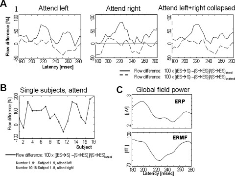
Figure 4.

A: The enhanced cumulated information flow from contralateral extrastriate (ES) to striate (S) cortical areas, related to the flow (S→ES) under the “attend” and the “neutral” condition. Both the EEG and MEG data were included when deriving the flow values (see methods section for details). All data were low pass filtered at 24 Hz cut off frequency. B: Single subject data of the contralateral flow form extrastriate to striate versus the flow from striate to extrastriate cortex. Same normalization as in subfigure A. C: Global field power for the event related potentials (ERP) and the event related magnetic fields (ERMF). The data reflect the attend left + right collapsed condition.
