
Figure 1.
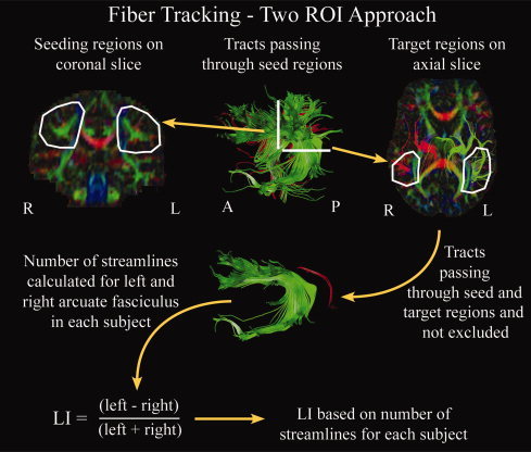
Schematic of tracking methods used to delineate the arcuate fasciculus. A manual two region‐of‐interest approach was used in which a seeding region was drawn on a coronal slice and a target region outlined on an axial slice in each hemisphere for each subject. Exclusion regions were used as needed to eliminate spurious streamlines, and were often needed in the area of the internal capsule. Tracts passing through both the seeding and target regions, without passing through exclusion regions, were retained for calculation of the lateralization index (LI). LI was calculated for each subject based on the number of streamlines in each hemisphere. [Color figure can be viewed in the online issue, which is available at www.interscience.wiley.com.]