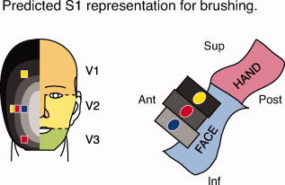
Figure 1.

Hypothesized segmental representation model of the face in S1. Predicted somatotopic arrangement with multiple stimulus sites based on the “onion‐skin” model, which refers to the onion‐like segmental pattern of facial representation. On the schematic of the face, the yellow, blue, and red squares represent stimulation sites along the superior–inferior and medio‐lateral axes. The orange, yellow, and green face regions represent areas innervated by the ophthalmic (V1), maxillary (V2), and mandibular (V3) divisions of the trigeminal nerve, respectively. Concentric ovals in shades from light gray (rostral) to black (caudal) represent hypothesized rostro‐caudal segmental divisions of the face. The cartoon to the right represents a sagittal view of the primary somatosensory cortex (S1) in the left hemisphere (contralateral to the stimuli). Prior data suggest that different regions of the face are represented segmentally in S1. The gray rectangles portray the cortical fields that correspond to the different segmental divisions on the face illustration. For example, the blue stimulation site is located most rostral on the face, and is represented in a relatively inferior location in S1. Note that stimulus sites along the medio‐lateral surface of the face are within the same division of the trigeminal nerve (V2), but are predicted to have a segmental representation in S1. Stimulation sites may also span the same segmental divisions, as shown by stimulation sites colored identically. Adapted from DaSilva et al [2002].
