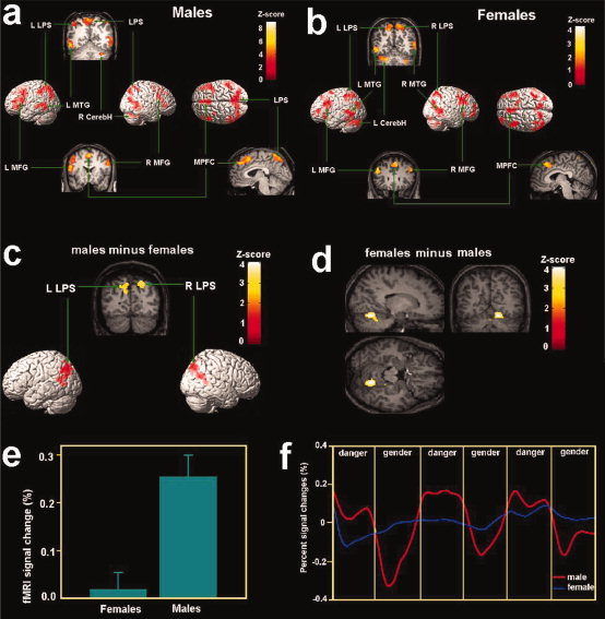
Figure 2.

Brain activation associated with danger detection in Experiment 1 shown in the random effect analysis. (a) Brain activations associated with danger detection in males; (b) brain activations associated with danger detection in females; (c) stronger posterior parietal activity was observed in the posterior parietal cortex in males than females during danger detection; (d) stronger cerebellar activity was observed in the cerebellum in females than males during danger detection; (e) percent signal changes of activity in the right parietal cortex was larger for males than females; (f) The time course of signal changes of the ROI in the right parietal cortex. The x‐axis of the time course is for the whole session of Experiment 1. Each local segment of the time course contains one 60‐s epoch in the scan corresponding to danger detection or gender identification. The time courses were averaged from raw fMRI signals. LPS = the superior parietal lobe, MFG = middle frontal gyrus; MPFC = medial prefrontal cortex, MTG = middle temporal gyrus; CerebH = cerebellum.
