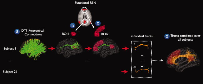
Figure 1.

Structural connections between functionally connected resting‐state network regions. The existence of possible anatomical white matter tracts between functionally connected resting‐state network (RSN) regions was examined in a 3‐step procedure. First, for each individual, the DTI data was used to reconstruct the white matter tracts of the human brain, called fibers or tracts (step a). Second, for each of the nine functionally connected RSNs, a first region of interest (ROI) was selected and from the total collection of fibers, the tracts that reached the first ROI were selected (step b). Third, a second ROI was retrieved from the RSN clustermap and from the resulting tracts of the previous step, only the tracts that reached this second ROI were selected (step c). For illustration purposes, the individual interconnecting tracts were normalized to match standard space (MNI 305) and combined over the group of subjects. The combined tracts represented the existence of a direct anatomical white matter connection between the functionally connected regions of a known resting‐state network.
