
Figure 1.
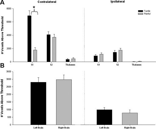
Extent of tactile‐ and pain‐evoked activations (thresholded method). (A) The mean (± SE) number of voxels (across all subjects) activated during tactile (filled bars) or painful stimulation (gray bars) is displayed for each brain region. There was a significant interaction between brain region and type of stimulation; post hoc testing revealed the presence of significantly more activated voxels in the contralateral S1 during tactile stimulation than during painful stimulation (P < 0.001; t = 6.23; denoted by asterisk), but there were no significant differences in the number of voxels activated by tactile versus painful stimulation in the contralateral S2 and thalamus or in ipsilateral regions (P > 0.05). (B) The mean (± SE) number of voxels activated over all left (filled bars) or right (gray bars) brain regions are displayed. Three‐way ANOVAs indicated no significant main effect of stimulation side. (S1, primary somatosensory cortex; S2, secondary somatosensory cortex).