
Figure 4.
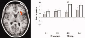
Differences in brain activation between women and men in left anterior insula. Increased activation in men was found for I‐3 (mildly painful). Statistical parametric maps are overlaid on a T1 scan (radiological convention: left = right, z = 8). The plots show parameter estimates for each rating (mean ± standard error for maximally activated voxel), indicating also a tendency of increased activation in men compared to women for I‐4 (moderately painful). The asterisk indicates significant differences between sexes.