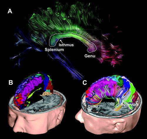
Figure 2.

Callosal fiber parcellation of a subject using cortical gray matter parcellation. Approximately 64 gray matter parcellations from the structural T1 image were used to parcellate the white matter fiber bundles. Panel A shows a cross‐section of callosal fibers in the mid‐sagittal plane. The cross‐section is displayed with continuous colors irrelevant to the cortical gray matter parcellation. The callosal fiber colors in panel A were assigned by their similarity to neighboring fibers, i.e., similar directional fibers have similar colors. Panel B shows cortical parcellation that was used to parcellate callosal fibers according to their connection to the cortical lobes. Panel C shows the parcellated callosal fibers color‐coded according to the assigned colors of their interconnecting cortical lobes.
