
Figure 1.
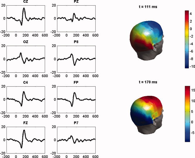
(a) Left: Average (over 76 trials) of the difference face—scrambled face EEG data for the channels CZ, PZ, IZ, P8, C4, FPZ, FZ, and P7, plotted against time t(ms). There is a negative EEG peak around t = 111 ms for the electrodes CZ, PZ, C4 in the temporal region, and FPZ and FZ in the frontal region, followed by a positive peak around 170 ms. The reverse happens in the occipito‐temporal channels OZ, P8, and P7 (the so‐called N170 component). (b) Right: the scalp topographic distribution of the 128 channels at the time instants t = 111 ms and t = 170 ms. [Color figure can be viewed in the online issue, which is available at www.interscience.wiley.com.]