
Figure 3.
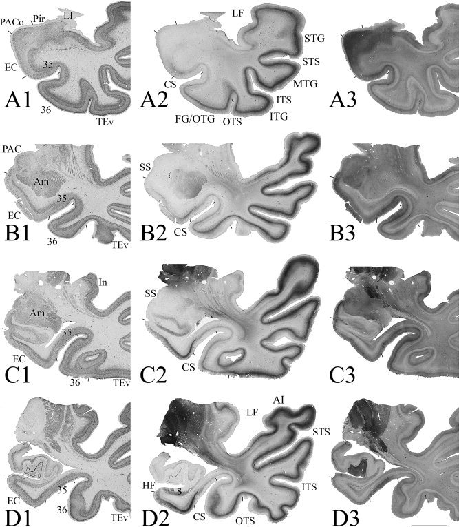
Photomicrographs of sequential coronal sections through four different anterior–posterior (AP) levels (A–D) of the MTL from a normal case show the main features of areas 35 and 36 in comparison with adjoining areas. This case has a Type I CS (deep CS) and no RS. At each AP level three adjacent sections were shown and they were stained for NeuN (A1‐D1), PV (A2‐D2), and CB (A3‐D3), respectively. Borders of areas 35 and 36 were defined by a combined analysis of the different markers. Examples of areas 35 and 36 at higher magnifications from this case were shown in Figure 4. For abbreviations, see list. Bar: 1 cm.