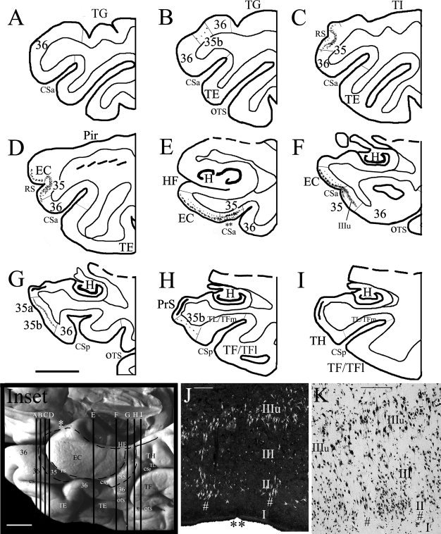
Figure 7.

Localization and topography of human area 35 in a normal aging case with a Type II CS (deeper CS) and a deeper RS, as determined with Thioflavin‐S and Nissl stains in sequential coronal sections. Area 35 and its adjoining areas were outlined in the inset, where the location of the LI (indicated by two white asterisks) and sections (A)–(I) were marked. (A–I) The location of large NFT+ cells at different AP levels in areas 35 and the EC. The microphotograph of the area 35 in (E) was shown in (J) (the double black asterisks in E and J mark corresponding region). Note that in the most anterior and posterior coronal sections (B, H), large NFT+ pyramidal cells can be observed only in the unique layer IIIu of area 35b but not in the adjoining areas 36, TH, and TL/TFm. (J, K) Microphotographs of the sections from area 35 stained with Thioflavin‐S (J) and NeuN (K) show tau pathology (J) and normal cytology (K) of area 35. Note the early tau lesion in the large pyramidal neurons in layer IIIu and in large‐celled vertical columns (indicated by #) of the superficial layers. For abbreviations, see list. Scale bars: 1 cm in (A)–(I) and inset; 300 μm in J; 200 μm in K.
