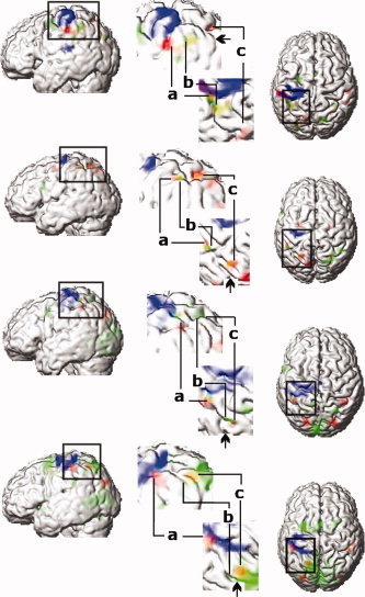
Figure 3.

Activated regions in four individual subjects, (top to bottom). Significant activations for surface orientation vs. color discrimination (red) (P < 0.001 uncorrected), for the motor response of the left index and middle finger (blue) (P < 0.05 corrected) and for delayed period for saccadic eye movements (green) (P < 0.05 corrected within fROI). Orange color indicates overlap between the areas of surface orientation discrimination and delay period prior to saccadic eye movements (Sub 9). First column, rendered brain viewed from left. Central column, an enlarged view of the areas around the IPS viewed from the right and from above. Right column, rendered brain viewed from above. In the enlarged view, activations during orientation discrimination (a, AIP; c, CIP) are located in anterior and posterior part of IPS. Activations related to the delay period prior to saccadic eye movement (b, middle part of IPS) are located between AIP and CIP. Arrows in enlarged view indicate border of parietal and occipital lobe in IPS.]
