
Figure 3.
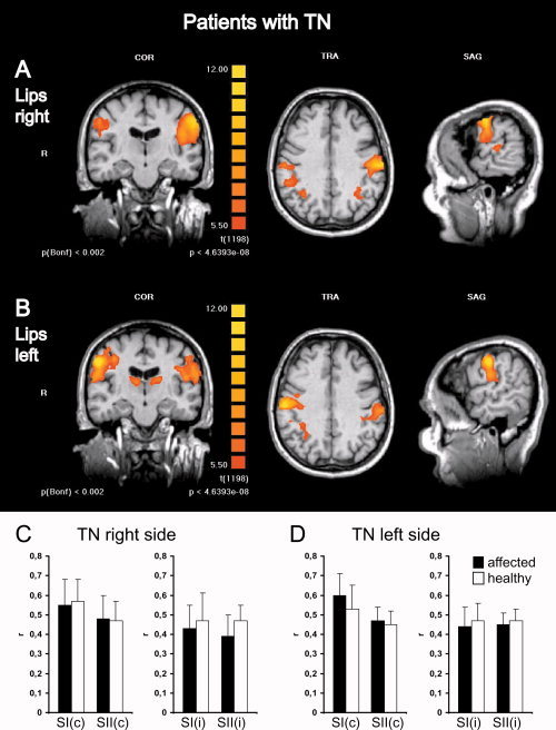
Somatosensory activations in patients with TN show no differences depending on stimulation of the affected or unaffected body side. Group level fMRI of bilateral somatosensory activations in the postcentral gyrus and parietal operculum in patients with TN on the right side are shown for stimulation of the right (A) and left (B) lips. To conserve anatomical details, group level BOLD‐activation maps (n = 11) were overlaid onto anatomical images of one individual subject. Left panels show a coronal view (COR) through the plane of the postcentral gyrus, middle panels a transversal view (TRA) through the plane of SI activations and left panels a sagittal view (SAG) through the plane of contralateral activations. R, right, L, left. For display of group data (different from Fig. 2) colors depicting the t‐statistic with uncorrected and Bonferoni corrected P‐values were chosen to facilitate comparison with other studies where mostly t‐values are presented. Each activation map had a false discovery rate (FDR) < 0.001. Statistical thresholds were deliberately kept equal to allow comparison of activation maps. No major differences in activation levels can be observed depending on the stimulation side. Note bilateral thalamic activations in (B) (left panel) and coactivations in the superior parietal lobules in (A) and (B) (middle panel). Histograms depict quantitative analyses of r‐values (mean ± SD) comparing SI and SII activations elicited by lip stimulation on the affected (black bars) or the healthy side (white bars) for patients with TN on the right side (C) and left side (D) separately. c, contralateral; i, Ipsilateral; r, correlation of the measured BOLD‐signal to the applied hemodynamic reference function.