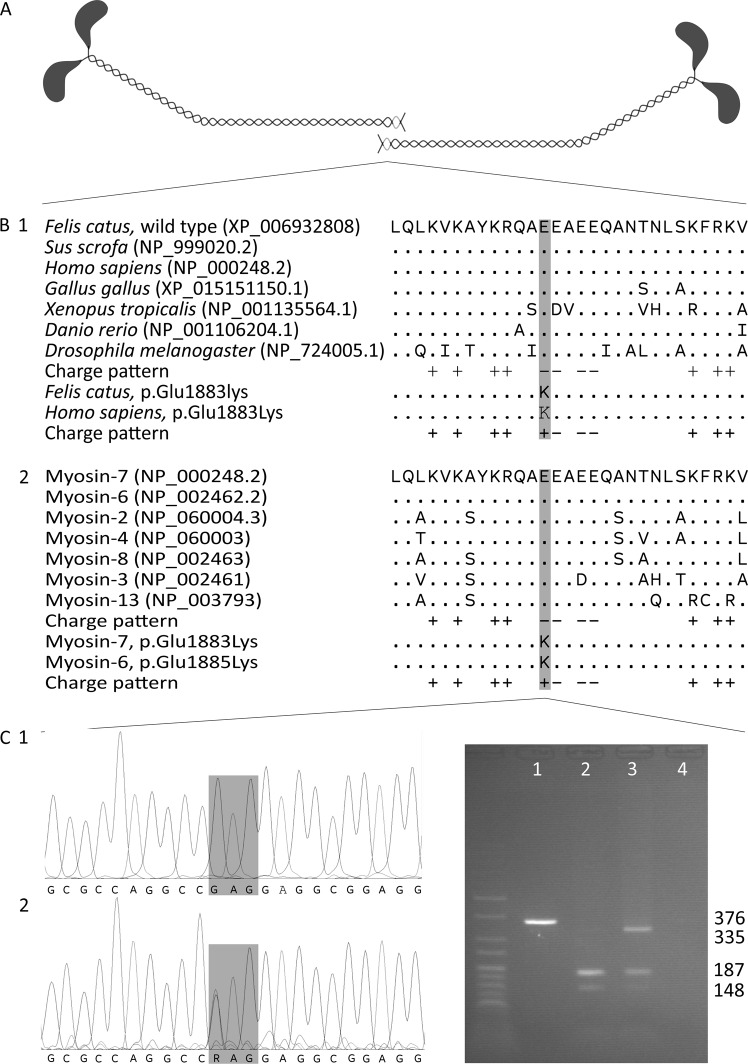
Fig. 1.
Position and evolutionary conservation of the ACD. a Tail-to-tail dimerisation of the coiled coils formed by myosin heavy chains. The ACD regions are shaded light-grey. b The amino acid sequence and charge pattern of the ACD in myosin-7 (myosin heavy chain for Drosophila) orthologues across species (1) and skeletal and cardiac muscle myosin heavy chain paralogues in humans (2). Accession numbers are given in parentheses and identical amino acids are depicted as “.”. The variants cause the substitution of a conserved, negatively charged glutamic acid residue by a positively charged lysine residue. c Chromatograms of a homozygous wild-type cat (1) and the heterozygous case (2). The variant changes codon 1883 from GAG to AAG. d Agarose gel electrophoresis of an uncleaved amplicon (1), 376 bp long, the cleaved amplicons of a homozygous wild-type cat (2) and the heterozygote case (3) and a negative control (4). The amplicon is always cleaved at an internal cleavage site in a large fragment of 335 bp and a small fragment of 41 bp. The wild-type large fragment contains a second recognition site and is cleaved in fragments of 187 and 148 bp, while the variant large fragment does not contain a recognition site and remains intact. Two percent agarose gel with HyperLadder V

