
Figure 4.
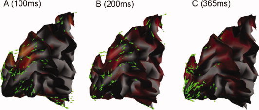
Sagittal views of the posterior portion of white‐grey matter interface the left mesial occipital lobe of one subject during the early response of the expanding rings protocol: the optical flow vector field is shown with green arrows at three different time latencies. The corresponding MEG current source density is shown in a light red shade.