
Figure 1.
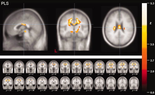
Results of the whole brain‐based analysis of the PLS patients. Local maximum of decreased FA values (thresholded at P < 0.05, corrected for multiple comparisons) in projections to the midbody of the corpus callosum (CC) in PLS patients in a sagittal, coronar and axial view (upper panel). The coronar multi‐slice view (lower panel) exhibits widespread clusters of reduced FA values within subcortical WM areas of mainly the motor system including the CST and the CC as well as the projectional fiber system of the upper brain stem and the frontal lobe. The significance level (Z‐score) is indicated by color temperature according to the scale (L = left).