
Figure 1.
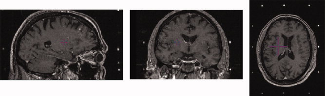
Example of pre‐operative MRI volume with headframe affixed to patient. Left: Sagittal view. Middle: Coronal view. Right: Axial view. Image volume shows lack of contrast in the subcortical nuclei. Surgical targeting is extremely difficult in these nuclei as a result. [Color figure can be viewed in the online issue, which is available at www.interscience.wiley.com.]