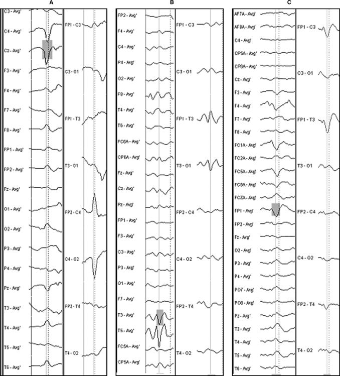
Figure 1.

Example of EEG selections (vertical line) for (A) PRS, (B) Left TS, and (C) Left FST. The left columns show the montage used for analysis (mean reference), the right columns show the classical bipolar montage used for clinical interpretation.
