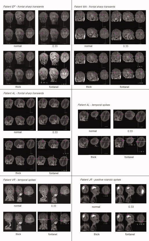
Figure 3.

Dipole fit results on averaged events obtained with the four head models, projection to 3D MRI. From top to bottom and left to right : E.F.'s frontal sharp transients (left FST above, right FST below), M.A.'s frontal sharp transients, A.L.'s frontal sharp transients, A.L.'s temporal spikes, V.R.'s temporal spikes and J.R.'s positive rolandic spikes. Patient A.L.'s MRI is showing an important ventricular dilatation, patient J.R.'s a Periventricular Leucomalacia with ventricular dilatation.
