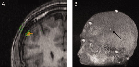
Figure 2.

A: In this coronal slice of a MRI image, the orange spot represents the DLPFC on the cortex (DLPFCC) and the green squares are voxels on the iso‐surface with a normal that passes through the DLPFCC in a distance less than 3 cm. Voxels (green squares) on surfaces other than the scalp (i.e., the internal side of the skull, and the interface grey matter‐CSF) were filtered out if they had a normal greater than 25° from the average normal. The mDLPFCS was defined as the intersection of the scalp with a line with the orientation of the average normal and passes through their average position. B: The position of mDLPFCS was superimposed on each individual's MRI for later co‐registration. Once mDLPFCS is co‐registered, this position is referred to as the eDLPFCS.
