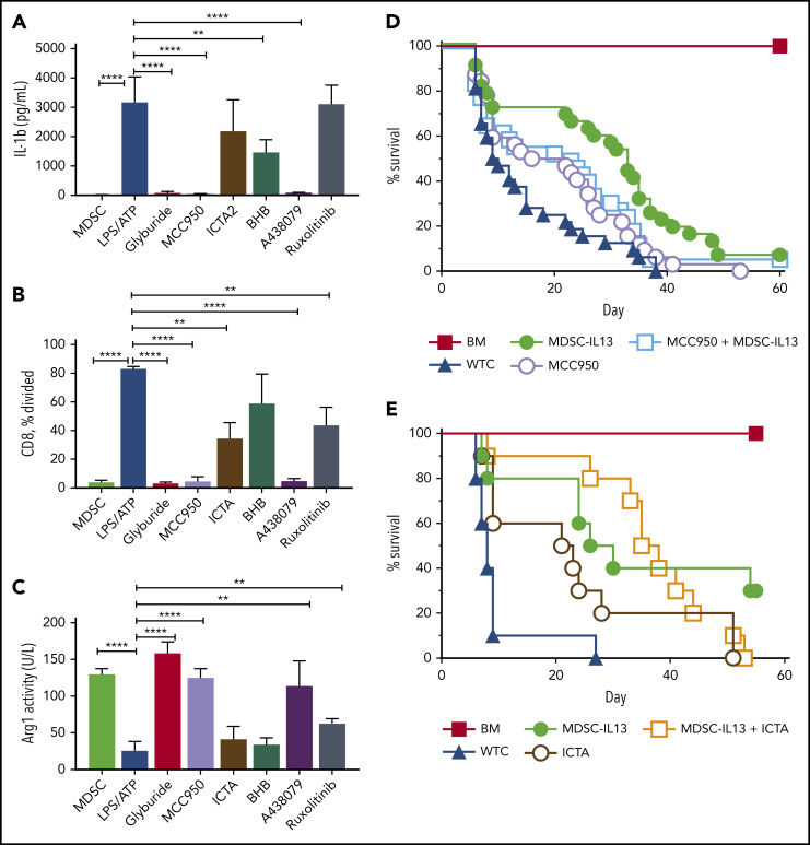
Figure 3.
NLRP3 inflammasome inhibition protects function of MDSC-IL13s. Cultured WT MDSC-IL13s were treated as indicated with reagents to inhibit inflammasome induction or myeloid maturation 1 hour before LPS plus ATP inflammasome induction: glyburide (100 µM), MCC950 (10 µM), ICTA2 (200 µg/ml), BHB (200 µM), A-438079 (25 µM), and ruxolitinib (1 µM). One hour after ATP stimulation, cultures were assayed as indicated. (A) Culture supernatants were assessed for IL-1β. (B) Harvested MDSCs were counted and plated for CFSE suppression assay at a 1:1 ratio with responding CD25-depleted whole T cells (WTCs); data represent frequency of CD8+ T cells undergoing ≥1 division. (C) MDSCs were washed, counted, and assayed for cell-associated arginase-1 activity. (A-C) In vitro data represent 3 replicates per condition and 2 independent experiments. (D) Kaplan-Meier survival curve for B6 > Balb/c GVHD animals given MDSC-IL13s (M13) and/or the NLRP3-specific inhibitor MCC950 (MCC) (50 mg/kg intraperitoneally) every other day starting at day −1 for 3 weeks. WTCs vs M13, P < .001; WTCs vs MCC, P = .0055; WTCs vs M13 plus MCC, P < .0001; M13 vs MCC, P = .0152; M13 vs M13 plus MCC, P = .4291; MCC vs MCC plus M13, P = .1226. Data represent combination of 3 independent experiments, n = 30 per group. (E) Kaplan-Meier survival curve for B6 > Balb/c GVHD animals given MDSC-IL13 (M13) and/or the NLRP3-specific inhibitor ICTA. WTCs vs M13, P = .0028; WTCs vs ICTA2, P = .0128; WTCs vs M13 plus ICTA, P = .0001; M13 or ICTA vs M13 plus ICTA, P = not significant. Data represent a single experiment, n = 10 per group. **P < .01, ****P < .0001.

