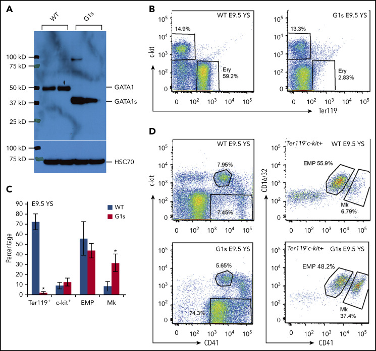
Figure 1.
Impaired yolk sac erythropoiesis in Gata1s embryos. (A) Western blot to detect the expression of GATA1 full length (GATA1) in WT embryos and the short isoform (GATA1s) in Gata1s mutant embryos (G1s). Cell lysates were extracted from E13.5 total fetal liver cells. Heat shock protein family A member 8 (HSC70) is shown as a loading control. (B) Flow cytometry assessment of the erythroid (Ery) population in E9.5 yolk sac (YS) using double-staining with antibodies against c-kit and Ter119. (C) Bar graph depicting mean (± SD) percentages of Ter119 positive, c-kit positive, EMP) and megakaryocyte (Mk) populations from yolk sacs of E9.5 WT and G1s as determined by flow cytometry. N ≥ 3. (D) Representative flow cytometry plots of EMPs and Mk stained with antibodies against c-kit, CD16/32 (FcγRIII and II), Ter119, and CD41. *P ≤ .05 (unpaired Student t test).

